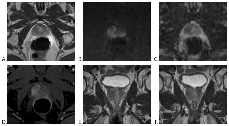
Figure 3.
63-year-old man with a PSA level of 14.32 ng/ml: mp-MRI with ERC, 7 weeks after positive prostate biopsy (Gleason score 3+3). A. Axial T2WI TSE, showing a homogeneous, hypointense mass in the peripheral zone of the right prostatic lobe. B. Axial DWI, C. ADC map and D. DCE-MRI showing mildly hyperintense signal on high b-value (b 1200) and markedly hypointense mass on ADC, with early enhancement. E. and F. Coronal T2WI TSE demonstrates broad capsular contact, bulging prostatic contour and breach of the capsule. All three readers reported a right prostatic mass with extraprostatic extension. The histopathological report after radical prostatectomy certified extracapsular extension (T3a).
PSA = prostate-specific antigen, mp-MRI = multiparametric MRI, ERC = endorectal coil, TSE = turbo spin echo.

