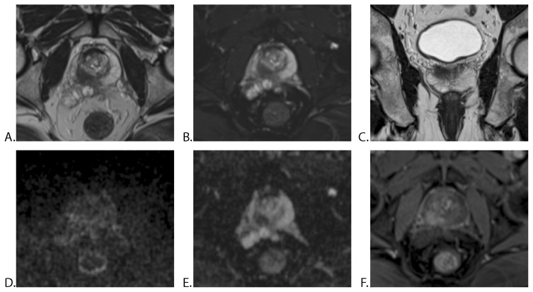
Figure 4.
57-year-old man with a PSA level of 9.0 ng/ml: mp-MRI without ERC (no prior prostate biopsy). A. Axial T2WI TSE, B. Axial T2WI fat-sat TSE and C. Coronal T2WI TSE showing a hypointense mass in the peripheral zone of the right prostatic lobe. D. Axial DWI image and E. ADC map demonstrates mildly hyperintense signal on high b-value (b 1200) and markedly hypointense mass on ADC, with early enhancement on F. DCE-MRI. All three radiologists reported a mass with extraprostatic extension and right seminal vesicle invasion. TRUS-guided biopsy performed after mp-MRI was positive for prostate cancer in the right gland with a Gleason score of 7 (3+4). Histopathological analysis after radical prostatectomy confirmed right seminal vesicle invasion by the prostatic adenocarcinoma (T3b).
PSA = prostate-specific antigen, mp-MRI = multiparametric MRI, ERC = endorectal coil, TSE = turbo spin echo, TRUS : transrectal ultrasonography.

