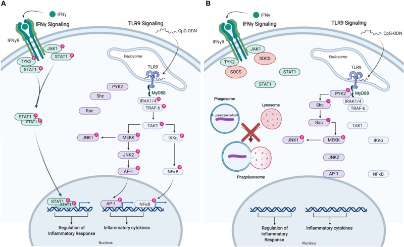
FIGURE 4.

Immune signaling pathways in bovine monocytes. Schematic representation of the IFNγ and TLR9 signaling pathways in response to exogenous stimuli and M. avium subsp. paratuberculosis infection. (A) Signaling pathways activated in uninfected bovine monocyte following stimulation with IFNγ, and TLR9 agonist CpG-ODN. Both pathways lead to the downstream phosphorylation of unique intermediates terminating with the translocation of distinct transcriptional activators into the nucleus to induce the expression of pro-inflammatory responses and cytokines. (B) M. paratuberculosis infection of bovine monocytes interferes with IFNγ signaling near the receptor by inducing the expression of SOCS, which disrupts JAK-STAT signaling. M. paratuberculosis infection also dysregulates TLR9 signaling by shunting the response toward the PYK2 pathway effectively blocking the induction of pro-inflammatory responses via MYD88. “P” designates protein in its phosphorylated state. Figure is adapted from Arsenault et al. (57, 58) and generated using BioRender. AP-1, adaptor protein complex 1; CpG-ODN, cytosine triphosphate-guanine triphosphate oligodeoxynucleotide; IFN, interferon; IKKα, inhibitor of nuclear factor kappa-B kinase subunit alpha; IRAK, interleukin-1 receptor-associated kinase 1; JAK, janus kinase; JNK, c-Jun N-terminal kinase; MEKK, MAPK/ERK kinase; MYD88, myeloid differentiation primary response protein; NFκB, nuclear factor kappa B; PYK, protein-tyrosine kinase; RAC, Ras-related C3 botulinum toxin substrate; SHC, Src homology 2 domain-containing-transforming protein; SOCS, suppressor of cytokine signaling; STAT, signal transducer and activator of transcription; TAK, TGF-beta-activated kinase; TRAF, TNF receptor-associated factor; TYK, non-receptor tyrosine protein kinase; TLR, Toll-like receptor.
