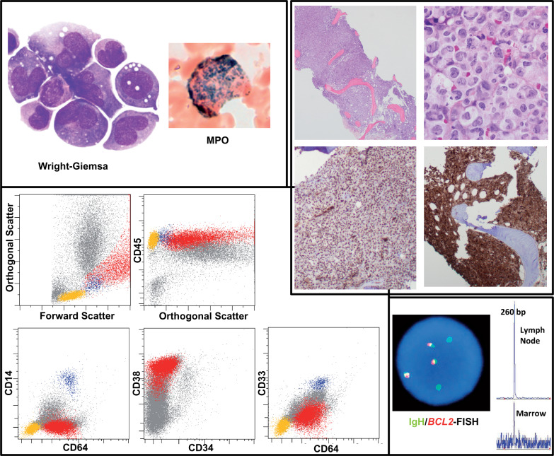
Figure 1.
Morphologic, immunohistochemical, flow cytometric, and genetic findings on bone-marrow specimen from our patient, a 52-year-old woman. Images at the top left depict tumor-cell morphologic characteristics on Wright-Giemsa–stained bone marrow smear preparations, with cytochemical staining for myeloperoxidase (MPO). The cluster of images at the top right shows hematoxylin-eosin (H&E)–stained sections of the bone-marrow core (showing 100% cellularity), with positive staining for PU.1 and lysozyme by immunohistochemistry. Dot plots on the bottom left demonstrate flow-cytometric findings (red: neoplastic population; blue: normal background monocytes, yellow: normal lymphocytes). Images at the bottom right depict the relatedness of the follicular lymphoma and myeloid leukemia: fluorescence in situ hybridization (FISH) positivity of tumor cells for the t(14;18) translocation involvingBCL2 (red: BCL2; green: immunoglobulin [Ig]H); and (2) presence of identical (260-bp) rearranged framework region 2 (FR2) IgH clonal peak in marrow and lymph-node specimens.CD indicates cluster of differentiation.

