Figure 1.
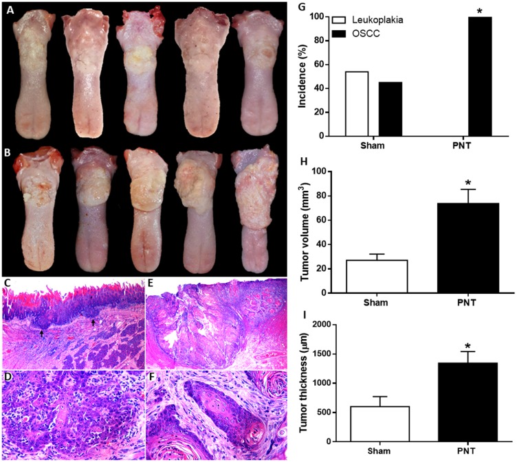
Clinical features of OSCCs derived from 4NQO treatment in sham (A) and PNT rats (B). (A) Small irregular white plates and discrete ulcerative lesions. (B) Extensive ulcerative lesions displaying reddish and yellowish-white areas. Histopathological features of tongue tumors from sham (C and D) and PNT rats (E and F) (H&E staining). (C) Well-differentiated OSCC (black arrows) (original magnification ×250). (D) Tumor cells showing hyperchromatism and dyskeratosis (original magnification ×400). (E) Extensive well-differentiated OSCC (original magnification ×250). (F) Islands of well-differentiated tumor cells with nuclear pleomorphism and keratin pearls (original magnification ×400). Occurrence and progression of OSCC in sham and PNT animals. (G) Chi-square test revealed that PNT rats had a higher OSCC occurrence than sham rats. (H) Student’s t-test showed that PNT group had increased tumor volume when compared to sham group. (I) PNT rats exhibited higher tumor thickness than sham-operated animals. (sham, n = 11; PNT, n = 12). Bars represent the mean ± SEM. * p < 0.05. The Figure 1E was spliced for joining together two images due to the extent of the tumor.

