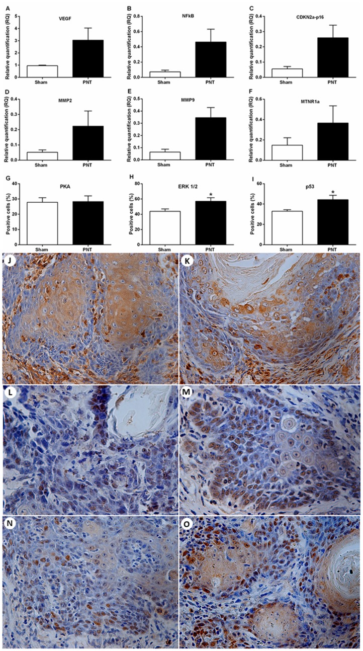
Figure 4. Expression of tumor progression-related genes and melatonin receptors and immunostaining for PKA, ERK1/2 and p53 in the OSCC microenvironment.
(A–F) Student’s t-test showed no differences between the groups concerning the mRNA expression for VEGF, NFkB, CDKN2A-p16, MMP2, MMP9 and MTNR1a in the tumor microenvironment. (G) Student’s t-test showed no statistical differences between both groups for PKA expression in the OSCCs. (H) PNT rats had increased tumor expression of ERK1/2 compared to sham animals. (I) PNT rats displayed higher tumor expression of nuclear p53 than sham rats. Immunoexpression of PKA (J and K), ERK1/2 (L and M) and p53 (N and O) in the OSCC invasion front from sham and PNT rats, respectively (original magnification ×400). Bars represent the mean ± SEM. * p < 0.05. (sham-PNT, n = 7; PNT, n = 9).

