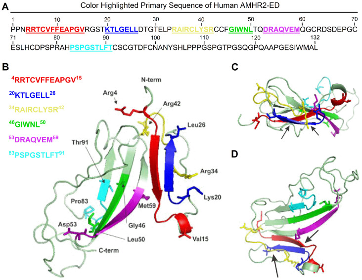
Figure 4. Molecular modeling of human AMHR2-ED.
Proposed binding sites for the cognate AMH ligand include the key natural AMH binding site I, 4RRTCVFFEAPGV15 represented in red in (A) the primary amino acid sequence and as a red beta sheet in (B) the ribbon model juxtaposed but antiparallel to the 20KTLGELL26 binding site of the 4D12G1 mAb depicted in blue in both the primary sequence and as a beta sheet in the ribbon model. Another strong AMH binding site II, 34RAIRCLYSR42 is represented by a long yellow loop also adjacent to the 20KTLGELL26 binding site of the 4D12G1 mAb. In contrast, the 53DRAQVEM59 binding site of the 12G4 mAb and its 3C23K humanized and glyco-engineered variant (a. k. a. GM102) is closer to the weaker tertiary and quaternary AMH binding sites 46GIWNL50 and 83PSPGSTLFT91, respectively. Arrows point to the juxtaposition of the blue 20KTLGELL26 binding site of the 4D12G1 mAb with the red AMH I binding site, 4RRTCVFFEAPGV15 and the yellow AMH binding site II 34RAIRCLYSR42 in (C) a top view of the ribbon model as well as in (D) a 90° rotational view.

