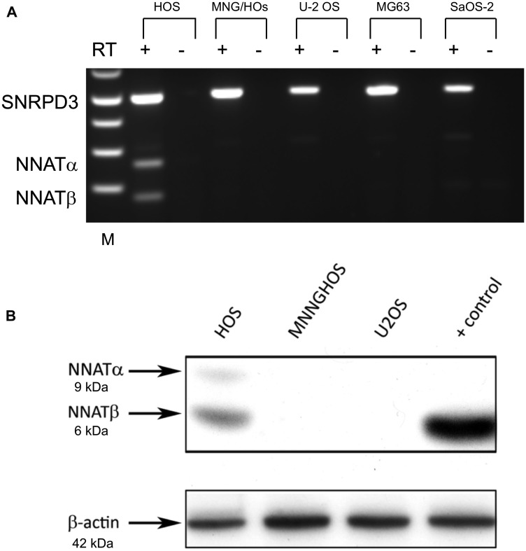
Figure 3. NNAT expression is detected in an osteosarcoma cell line exhibiting the normal imprinted hemimethylation pattern but not in cell lines exhibiting NNAT hypermethylation.
(A) NNAT mRNA expression in 5 osteosarcoma cell lines was assessed by semiquantitative endpoint RT-PCR. Total RNA was reverse transcribed and amplified with primers specific for NNAT or SNRPD3 coding sequences. Samples are (L to R): 1 kb ladder, HOS/reverse transcriptase (RT)+, HOS/RT–, MNNGHOS/RT+, MNNGHOS/RT-, U-2 OS/RT+, U-2 OS/RT–, MG-63/RT+, MG-63/RT–, Saos-2/RT+, Saos-2/RT–. The positions of SNRPD3, NNATα, and NNATβ bands are indicated. (B) NNAT protein expression in osteosarcoma cell lines was determined by immunoblot analysis of whole cell lysates. NNAT protein isoforms α and β are indicated. Cell lysate prepared from U-2 OS cells transiently transfected to express NNATβ was used as the positive control. Endogenous β-actin was detected to control for loading.

