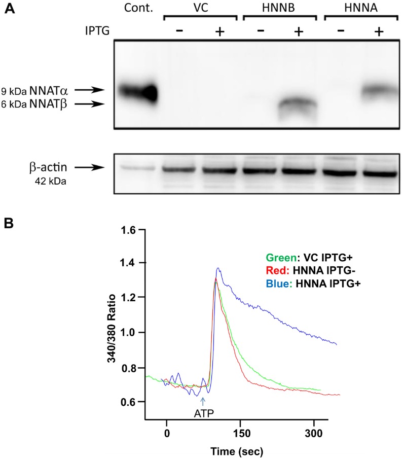
Figure 7. NNAT expression is associated with attenuated decay of [Ca2+]i following ATP-induced release of calcium stores in human osteosarcoma cells.
(A) MNNG/HOS cells were engineered for stable, IPTG-inducible expression of NNATα or NNATβ as described in Methods. NNATα (HNNA) and NNATβ (HNNB) expression was assayed by immunoblot in whole cell lysates of cells with and without IPTG treatment as indicated. VC represents empty vector control non-expressors. A lysate of MNNG/HOS cells transiently transfected to express NNATα was used as a positive control. (B) [Ca2+]i was measured by fluorescence imaging in fura-2/AM-loaded cells following ATP stimulation. HNNA cells were imaged with (blue tracing) and without IPTG (red tracing) induction of NNATα expression. VC cells are empty vector-control non-expressors imaged after IPTG treatment (green tracing). Shown are representative tracings from 3 replicate experiments.

