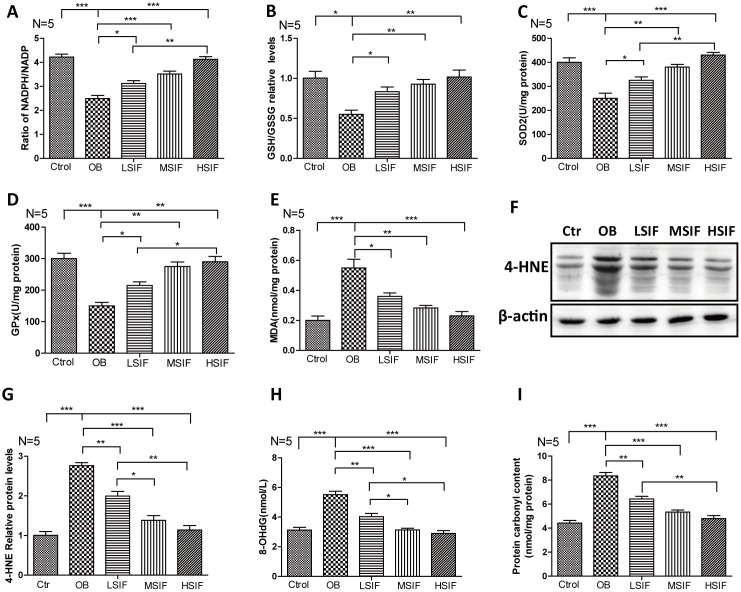
Figure 3.
Soy isoflavones reduce oxidative stress in hypothalamus of DIO male mice. (A) Quantification shows the NADPH/NADP ratio in hypothalamus of DIO mice fed with basal diets and the addition with different doses of soy isoflavones; (B) Quantification shows the GSH/GSSG ratio in hypothalamus of DIO mice fed with basal diets and the addition with different doses of soy isoflavones; (C) Quantification shows the SOD2 activity in hypothalamus of DIO mice fed with basal diets and the addition with different doses of soy isoflavones; (D) Quantification shows the GPx activity in hypothalamus of DIO mice fed with basal diets and the addition with different doses of soy isoflavones; (E) Quantification shows the MDA levels in hypothalamus of DIO mice fed with basal diets and the addition with different doses of soy isoflavones; (F, G) Western blots and quantification show the 4-HNE levels in hypothalamus of DIO mice fed with basal diets and the addition with different doses of soy isoflavones; (H) Quantification shows the 8-OHdG levels in hypothalamus of DIO mice fed with basal diets and the addition with different doses of soy isoflavones; (I) Quantification shows the protein carbonyl contents levels in hypothalamus of DIO mice fed with basal diets and the addition with different doses of soy isoflavones. * p < 0.05, ** p < 0.01, ***p < 0.001.

