Figure 1.
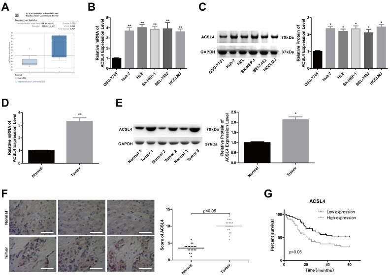
ACSL4 was overexpressed in HCC tissue samples and cells. (A) The Oncomine database was used to retrieve the different expression patterns of ACSL4 in HCC or normal liver. (B, C) The mRNA and protein content of ACSL4 in the human normal liver cell line QSG-7701 and the HCC cell lines Huh-7, HLE, SK-HEP-1, BEL-7402 and HCCLM3 were determined by RT-PCR and western blotting, respectively. (D, E) The mRNA and protein content of ACSL4 in HCC tissues and normal tissues were detected by RT-PCR and western blotting assays. (F) Immunohistochemistry was used to detect ACSL4 protein expression in HCC tissues and normal tissues (Scale bar = 100 μm). (G) Kaplan-Meier analysis of the relationship between ACSL4 expression and the overall survival of patients with HCC. (*P<0.05, **P<0.01).

