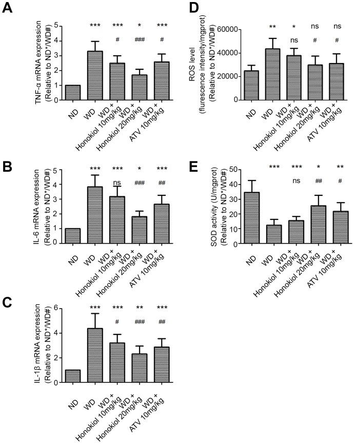
Figure 2.
Effect of honokiol on inflammatory response and oxidative stress in the carotid tissue of atherosclerotic mice. (A–C) The mRNA expression of TNF-α (A), IL-6 (B), and IL-1β (C) in carotid tissue was detected by real-time PCR. (n = 6; * P < 0.05, ** P < 0.01, *** P < 0.001, vs. the ND group. # P < 0.05, ## P < 0.01, ### P < 0.001, vs. the WD group; one-way ANOVA). (D, E) The ROS level (D) and SOD activity (E) in carotid tissue were detected by commercial kits in the indicated group. (n = 6; * P < 0.05, ** P < 0.01, *** P < 0.001, vs. the ND group. # P < 0.05, ## P < 0.01, ### P < 0.001, vs. the WD group; one-way ANOVA). TNF-α: Tumor necrosis factor-α; interleukin-6: IL-6; and interleukin-1β: IL-1β; ND: normal diet; WD: Western-type diet; ROS: reactive oxygen species; SOD: superoxide dismutase.

