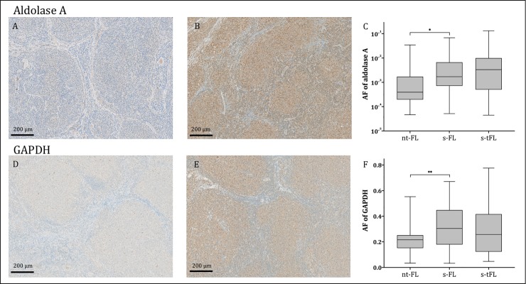
Fig 1. Predictive biomarker expression levels.
Representative images of tumor tissues stained for aldolase A (A-B) and GAPDH (D-E). A and D show tumor tissue from patients with no subsequent transformation (nt-FL), while images B and E show tumor tissue from the initial FL diagnosis, from a patient with subsequent transformation (s-FL). Area fractions of strong intensity aldolase A staining (C) and GAPDH (F) were significantly higher in FL diagnostic samples of patients with subsequent transformation (s-FL) compared with patients without (nt-FL); *p<0.001, **p<0.01. Two patients were not eligible for GAPDH IHC analysis and thus 39 nt-FL patients remained in this group (F). No significant difference in aldolase A and GAPDH expression was observed between FL diagnosis (s-FL samples) and time of transformation (s-tFL samples) (C and F). AF, area fraction; immunohistochemistry; FL, follicular lymphoma; FLIPI, follicular lymphoma international prognostic index; GAPDH, glyceraldehyde-3-phosphate dehydrogenase; HT, histological transformation; LDH, lactate dehydrogenase; PFS, progression free survival; TFS, transformation free survival.

