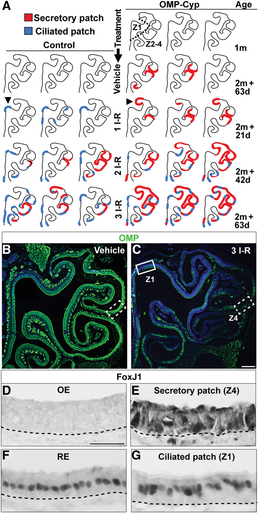
Figure 6.
Two types of metaplastic patches appear after methimazole injury or high-level transgenic expression of CYP26B1. A, Schematic illustrations of OE hemisections representing three OMP-Cyp mice at 1 month of age (1m). Also illustrated are OE hemisections of control and OMP-Cyp mice after one, two, and three I–R cycles according to the schedule outlined in Figure 4O. Red lines indicate patches with metaplastic secretory respiratory cells (secretory patch), whereas blue lines indicate patches with metaplastic ciliated respiratory cells (ciliated patch). Arrowheads indicate examples of OE regions without flanking RE where isolated patches within otherwise normal OE histologically were identified. B, C, Distribution of OMP+ OSNs (green) in control mice after vehicle (B) and after three I–R cycles (C) is shown. Nuclei are in blue. Indicated Z1 region (solid box) and Z4 region (dashed lined boxes) correspond to the regions analyzed in D, E, and G. D–G, Show FoxJ1 immunostaining in Z4 (D), a patch of secretory respiratory metaplasia in Z4 (E), RE (F), and a patch of ciliated respiratory metaplasia in Z1 patch (G). Scale bars: C, 300; D, 12.5 µm.

