Figure 7.
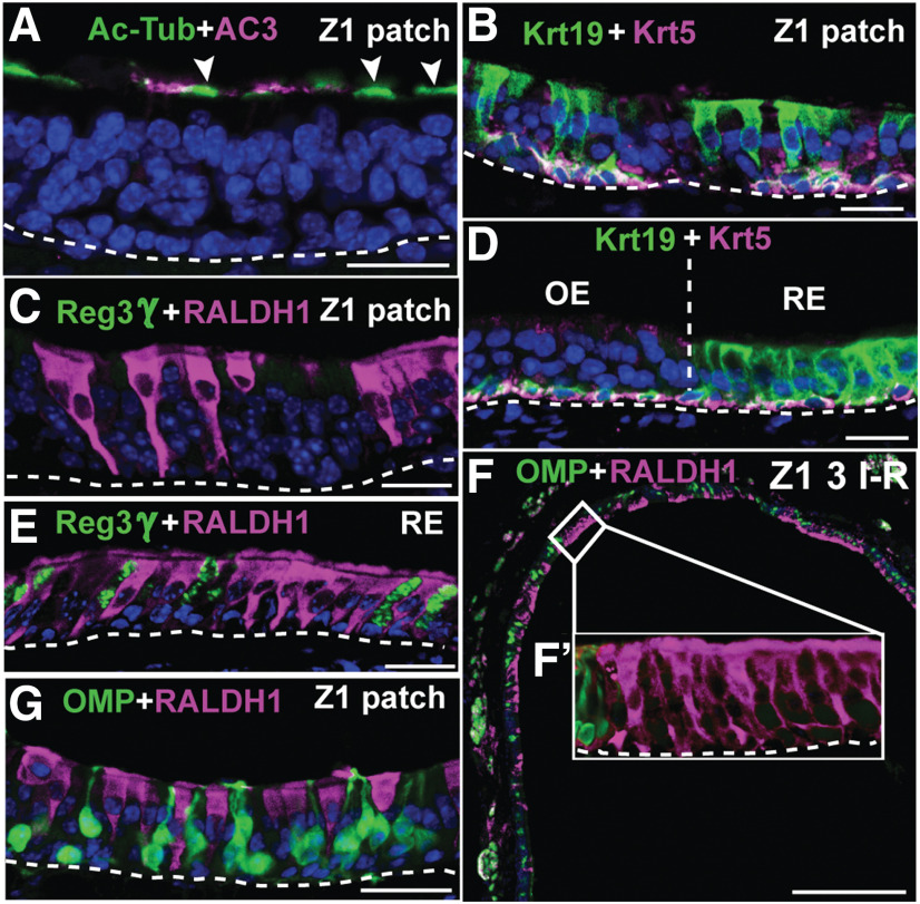
Metaplastic cells in Z1 are ciliated respiratory cells. A, Ac-Tub (green) and AC3 (magenta) analyses of a Z1 patch showing that Ac-Tub cilia (arrowheads) staining of the metaplastic cells does not overlap with OSN-specific AC3+ cilia staining. B, D, Krt19 and Krt5 immunofluorescence shows that both Z1 patch cells (B) and RE cells (D) express Krt19. C, E, Analysis for Reg3γ (green) and RALDH1 (magenta) shows that the Z1 patch cell type is RALDH1+/Reg3γ− (C), while both RALDH1+ and Reg3γ+ cells are present in RE (E). F, RALDH1+ ciliated respiratory cells (magenta) and OMP+ OSNs (green) along the dorsal nasal recess (i.e., Z1) of control mice after three I–R cycles. F´, Close-up of a patch with RALDH1+ respiratory cells that lacks OMP+ OSNs. G, A patch that has a mixture of RALDH1+ respiratory cells and OMP+ OSNs. Scale bars: A–E, G, 12.5 µm; F, 200 µm. Nuclei are in blue.

