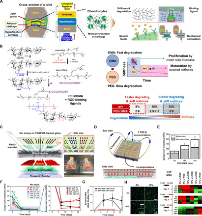
Fig. 1. Stem cell–based cartilage repair strategy is based on engineered multicomponent biomaterials and a combinatorial system for screening of combinatorial physical and biochemical cues in cartilage microenvironments.

(A) Schematic depicting key factors guiding chondrogenic differentiation of hMSCs. (B) Chemical synthesis scheme of hybrid of photo–cross-linkable PEG/OMA hydrogels. (C) Schematic showing six layers (i.e., TMSPA-treated glass slides with hydrogel arrays, caps to maintain gel height, main chamber, pistons with different heights, membrane with pillars, and pressure chamber) comprising the combinatorial high-throughput system. A representative photograph showing a combinatorial system for screening combinatorial cues (Photo credit: J.L., University of California, Los Angeles) (please also see fig. S1, F and G). Scale bar, 20 mm. (D) Schematic illustrations of cross-sectional view of the combinatorial system. (E) Compressive modulus of PEG/OMA hydrogels (8, 10, and 12% PEG/OMA) with or without cells on day 0 (n = 5) (One-way ANOVA with Tukey’s significant difference post hoc test; *P < 0.05 and ***P < 0.005 compared with 8% without cells). (F) Time profile of hydrogel degradation without compression for 21 days (n = 5). (One-way ANOVA with Tukey’s significant difference post hoc test; *P < 0.05 compared with 10% without cell group, **P < 0.05 compared with 12% with cell group, ***P < 0.005 compared with 8% with cells, and ****P < 0.005 compared with 12% without cell group at day 0.) (G) Relative DNA content of cells in PEG/OMA hydrogels with compositions of 8 and 12%, TGF-β1 (10 ng/ml), and RGD conjugation under 0 or 40% cyclic compression (n = 6). (H) Representative DAPI/F-actin images of hMSCs cultured for 7 days in 8 and 12% PEG/OMA gels with or without 40% cyclic compression and quantification of cell spreading at days 1, 3, and 7 (n = 3). Scale bar, 300 μm (inset: 100 μm).
