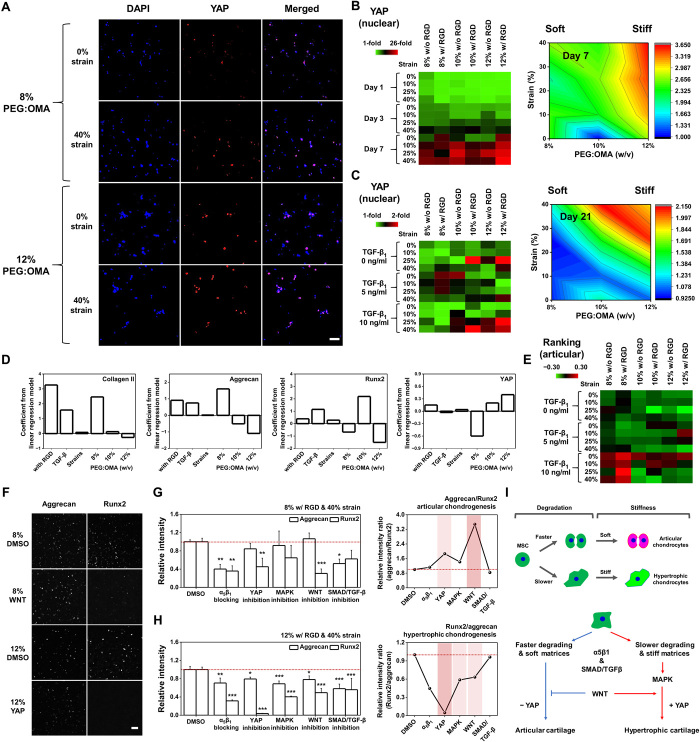
Fig. 3. Engineered multicomponent biomaterials regulate hypertrophic chondrogenesis of hMSCs through YAP-dependent mechanotransduction and WNT signaling pathway.
(A) Representative 3D confocal images of hMSCs cultured in different combinations of parameters for 7 days and stained with YAP. (B) Quantified heat map and surface plot of measured immunofluorescence nuclear intensity of YAP at days 1, 3, and 7 (n = 4). (C) Quantified heat map and surface plot of YAP for cells encapsulated in the hydrogel microarrays cultured with combinations of all the factors for 21 days (n = 4). (D) Plots of LR model coefficients across the parameters regulating expression levels of articular (collagen II and aggrecan) and hypertrophic (Runx2 and YAP) cartilage markers. (E) Quantified heat map of scores based on positive correlation of articular (collagen II and aggrecan) and negative correlation of hypertrophic (Runx2 and YAP) cartilage marker expression levels for the best chondrogenic metrics on day 21. (F) Representative 3D confocal images of cells stained with aggrecan and Runx2 when treated with an inhibitor of the WNT signaling pathway for the 8% PEG/OMA condition or of the YAP signaling pathway for the 12% PEG/OMA condition. Relative expression of aggrecan and Runx2 markers for cells cultured in (G) 8% PEG/OMA hydrogels with or without WNT inhibitor or (H) 12% PEG/OMA hydrogels with or without YAP inhibitor, demonstrating possible pathways that promote hypertrophy in articular-like or hypertrophic-like chondrogenesis (n = 4). (P values were obtained on the basis of one-way ANOVA with Tukey’s post hoc testing *P < 0.05, **P < 0.005, and ***P < 0.0005 compared with the DMSO condition.) (I) Proposed pathway for mechanical stimuli guiding articular or hypertrophic chondrogenesis of hMSCs. Scale bars, 100 μm.

