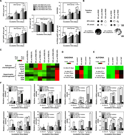
Fig. 4. hMSCs cultured in high-ranked conditions for the prevention of hypertrophic chondrogenesis show higher potency for cartilage repair in vitro and in vivo.

(A) Quantification of GAG/DNA and ALP/DNA for 21 days in culture of hMSCs in RGD-conjugated PEG/OMA gels supplemented with TGF-β1 under 40% compression compared with those cultured without compression (n = 6). (B) Toluidine blue O staining after treatment with threshold-based imaging analysis. (C) Heat map of real-time PCR gene expression results associated with articular or hypertrophic chondrogenic differentiation for cells cultured in the combinatorial bioreactor with modulation of mechanical stimuli for 21 days (n = 6 for Col2a1, ACAN, Sox9, and ALP & n = 3 for Col1a1, Col10a1, OCN, and OSX). Quantification of (D) GAG/DNA and (E) ALP/DNA for cells cultured for 21 days with or without WNT and YAP inhibitors and transplanted in vivo for 7 or 21 days after the in vitro pretreatment (n = 6). (F) Real-time PCR quantification of gene expression associated with chondrogenic (Col2a1, ACAN, and Sox9) or osteogenic (ALP, Col1a1, Col10a1, OCN, and OSX) differentiation for cells before and after transplantation in vivo (n = 3 to 5, please see fig. S9D). (*P < 0.05, **P < 0.005, and ***P < 0.0005 based on one-way ANOVA with Tukey’s post hoc testing). Scale bar, 100 μm.
