
Fig. 5 FISH analysis for tracking transplanted huMSCs in the endometrium.
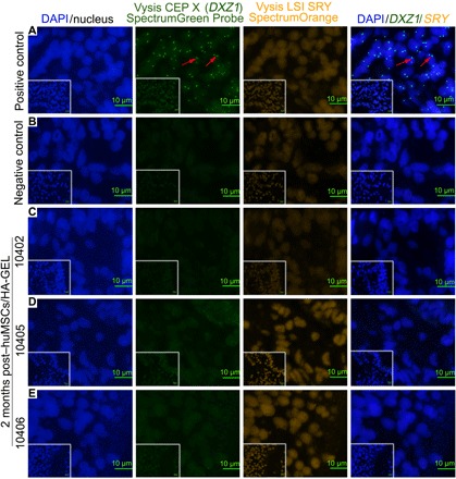
(A) Positive control, human endometrium (containing XX chromosomes); the red arrow indicates the green signal for Vysis CEP X (DXZ1). (B) Endometrial localization of huMSCs in the HA-GEL transplantation group (negative control). (C to E) Distribution of huMSCs in endometria 2 months after huMSCs/HA-GEL co-transplantation. Double/single-labeled staining (orange/green signal or just green signal) cells were defined as huMSCs. For details on 10402, 10405, and 10406, see table S3. Inserted overview pictures show a lower magnification.