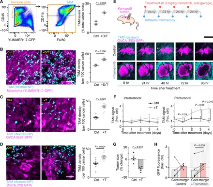
Fig. 2. MAPKi increases TAMs in BRAF-mutant melanoma and OVCA mouse models.

(A) YUMMER1.7 melanoma tumors were excised and analyzed by flow cytometry for TAMs (GFP− CD45+ CD11b+ F4/80+), 24 hours after three daily doses of dabrafenib (30 mg/kg) and trametinib (0.3 mg/kg) (+D/T; n ≥ 3). (B to D) Representative images (left) and quantification (right) of dextran-NP+ TAMs (cyan) within GFP-labeled (magenta) (B) YUMMER1.7, (C) intraperitoneally disseminated ES2 OVCA, or (D) intraperitoneally disseminated PtD OVCA tumors. At least one tumor each from n = 3 nu/nu mice per group was excised 24 hours after three daily doses of MAPKi (+T trametinib alone; two-tailed t test). Ctrl, control. Scale bars, 50 μm. (E to H) Schematic depicting daily imaging and trametinib treatment schedule for mice bearing ES2 xenograft tumors implanted in dorsal skin-fold window chambers (top). Representative intravital microscopy of OVCA and TAMs (bottom) (E) and their quantification (F) to (H) are shown (n ≥ 4 nu/nu mice per group, two-tailed t test). Scale bar, 250 μm. Tumor size and fluorescence were compared at 0 and 96 hours. Data are means ± SEM for all.
