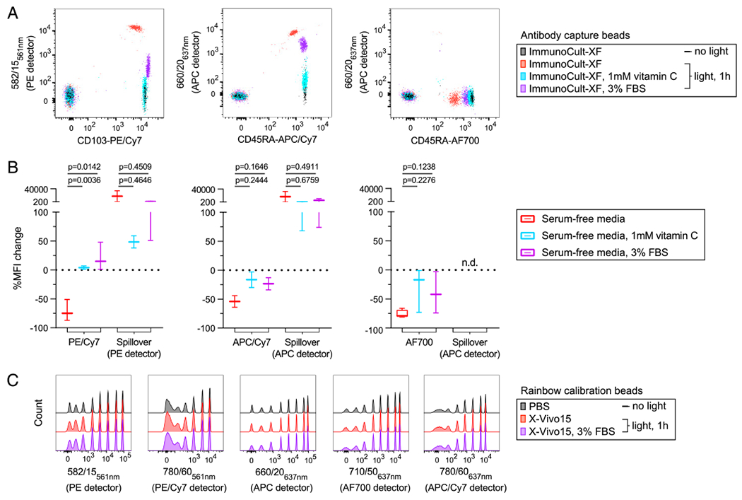
FIGURE 3. Serum and vitamin C limit light-initiated fluorochrome degradation in formulated serum-free media.

(A and B) Ab capture beads single stained with the indicated conjugates were resuspended in formulated serum-free media, either ImmunoCult-XF or X-Vivo15. (A) Fluorescence signal and spillover into adjacent detectors was measured immediately (no light) and again following exposure to ambient fluorescent light (light, 1 h) with or without either vitamin C or FBS. (B) A statistical comparison of the percentage of MFI change following light exposure is shown for data pooled from stained cells and Ab capture beads resuspended in either ImmunoCult-XF or X-Vivo15 serum-free media. (C) Rainbow calibration eight-peak beads were placed in PBS or formulated serum-free media, X-Vivo15 with or without FBS. Fluorescence signal was measured before and after light exposure as in (A) and for the same five detectors. Detector bandpass filter and laser line are shown. Significance was determined by ordinary one-way ANOVA with Dunnett posttest for multiple comparisons. Data are representative of at least two independent experiments. n.d., not detectable.
