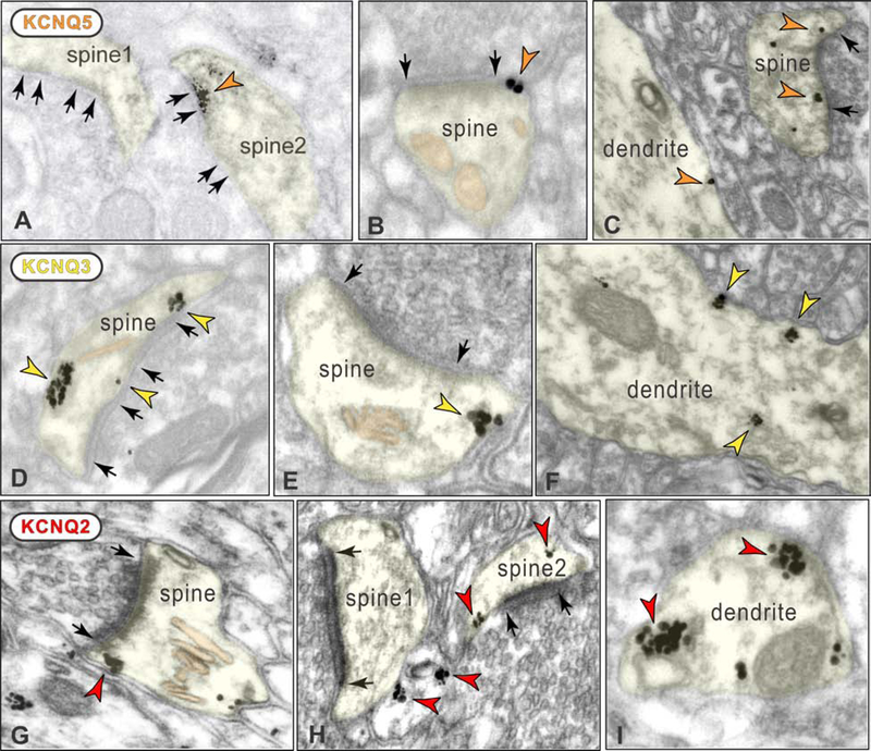
Figure 6. ImmunoEM localization of KCNQ isoforms on spines and dendrites in layer III dlPFC.
A-C. KCNQ5 localization on spines and dendrites. Panel A shows DAB KCNQ5 immunolabeling within the PSD in a perforated, asymmetric (glutamate-like) synapse (spine2); unlabeled synapses in spine1 and spine2 are indicated for comparison. Panel B shows immunogold KCNQ5 labeling immediately next to the PSD (peri-synaptic) in a spine. Panel C shows KCNQ5 immunogold peri-synaptic in a spine, and on the plasma membrane of a dendrite. D-F. KCNQ3 localization on spines and dendrites. Panel D shows KCNQ3 immunogold labeling in a spine with a perforated, asymmetric synapse. Panel E shows extrasynaptic KCNQ3 immunogold labeling in a spine receiving a glutamate-like asymmetric synapse. Panel F shows KCNQ3 immunogold labeling on the plasma membrane and within a dendrite. G-I. KCNQ2 localization on spines and dendrites. Panels G and H show KCNQ2 immunogold labeling in spines near synapses and at extrasynaptic locations. Panel H also shows label in an unidentified, likely glial profile. Panel I shows KCNQ2 immunogold labeling on the plasma membrane of a dendrite. Black arrows delineate synapses. The calcium-storing spine apparatus is pseudo-colored pink in panels B, D-E and G.

