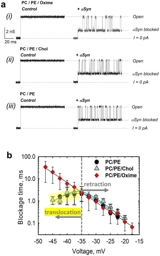
Fig. 4.

Olesoxime prevents αSyn translocation through reconstituted VDAC. a Representative ion current records of the single VDAC channel reconstituted into a planar lipid bilayer formed from (i) PC/PE (1:4 w/w) mixture with 5% (w/w) of olesoxime (PC/PE/Oxime) before (left column) and after (right column) addition of 50 nM of αSyn to the cis compartment were obtained on the same VDAC channel. The characteristic blockages of VDAC conductance induced by αSyn are seen in trace in the right column. Traces (ii) and (iii) were obtained in the membrane composed of PC/PE (1:4) with 5% (w/w) cholesterol (PC/PE/Chol) (traces ii) and PC/PE (1:4) (traces iii) before (left column) and after addition of 50 nM αSyn (right column). All records were taken at -40 mV applied voltage. The membrane-bathing solutions contained 1 M KCl buffered with 5 mM HEPES at pH 7.4. Dashed lines indicate VDAC open and αSyn-blocked states and zero current. All current records were smoothed with a 5 kHz low-pass Bessel digital filter using pClamp 10.3. b Voltage dependences of mean blockage times of αSyn-induced blockages of VDAC reconstituted into planar membranes made of PC/PE (1:4) with 5% (w/w) of olesoxime or cholesterol or in pure PC/PE (1:4) membranes. The increase of blockage time with absolute amplitude of the applied voltage corresponds to the pore blockage regime; a decrease of blockage time with voltage amplitude (highlighted in yellow for PE/PC and PE/PC/Chol membranes) corresponds to regime of αSyn translocation through the pore. In the presence of olesoxime, translocation of αSyn is not observed at applied voltages up to − 48 mV, while in PC/PE membranes with or without cholesterol, the translocation regime starts at − 35 mV. Data points and error bars represent the mean and SD for 4–6 independent experiments
