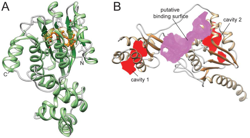
Figure 7– Structures of eye-specific retinoid-binding proteins.
(A) CRALBP in complex with 11-cis-retinaldehyde (PDB # 3HY5). The location of the retinoid molecule within the binding pocket is shown in orange. (B) The structure of module 2 from Xenopus laevis IRBP (PDB #1J7X). Two cavities that represent binding sites are marked in red, whereas a lipophilic hinge region is colored purple. The CavityPlus server was used to identify the intramolecular cavities and binding sites [58].

