Fig. 1.
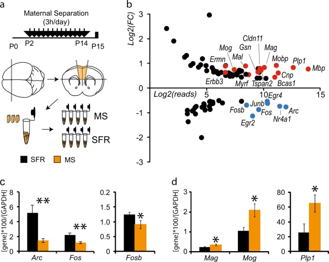
Analysis of differentially expressed genes in the medial prefrontal cortex (mPFC) of P15 animals exposed to maternal separation highlights myelin ensheathment and immediate early genes. a Schematic presentation of the experimental paradigm of the maternal separation (MS pups) versus standard facility (SFR pups) protocols and the dissection of mPFC at P15 to generate four samples per condition, each including three unilateral mPFC. b Graph shows with a log2 scale the fold change (FC) of expression induced by maternal separation according to the number of reads detected in control samples for the genes with q < 0.05 and FC > 1.3 or FC < 0.7. Myelin-ensheathment genes are highlighted in red, and immediate early genes in blue. c, d Graphs show the quantitative PCR (qPCR) results obtained in a second experimental cohort to determine the expression levels of IEG (C, n = 7 SFR animals and n = 6 MS) and genes related to myelin ensheathment (D, n = 6 SFR animals and 9 MS). *p < 0.05, **p < 0.01

