Fig. 2.
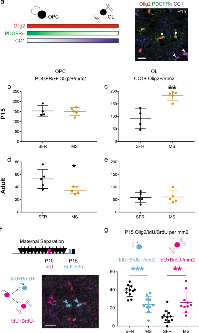
Maternal separation induces precocious differentiation of oligodendrocytes in the developing medial prefrontal cortex (mPFC). a The scheme illustrates the specificity of the oligodendrocyte lineage markers using triple immunostaining. Oligodendrocyte progenitor cells (OPCs) express Olig2 and high levels of PDGFRα while mature oligodendrocytes (OLs) express Olig2 and high levels of the CC1 antigen. Scale bar represents 20 µm. b–e Graphs show the density of OPCs (b, d) and OLs (c, e) in the P15 (b, c n = 5 animals per conditions) and adult (d, e n = 5 animals per condition) mPFCs of SFR and MS animals. f Scheme shows the protocol of sequential labelling of OPCs using IdU injections at P10 and BrdU injections at P15 to differentiate the fraction of P10 OPCs that are still cycling at P15 (blue, Olig2+IdU+BrdU+) from those which have started differentiation by then (purple, Olig2+IdU+BrdU−). g Graph shows a decresased fraction of cycling OPCs and an increased fraction of differentiated OPCs in P15 mPFC of MS animals as compared to SFR animals (n = 11 SFR animals and 10 MS animals). *p < 0.05, **p < 0.01, ***p < 0.005

