Fig. 3.
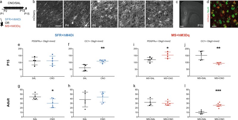
Neuronal activity bi-directionally controls oligodendrocytes differentiation in the developing medial prefrontal cortex (mPFC). a Scheme illustrates the experimental paradigms of the chronic treatment with CNO or SAL solutions from P2 to P14 after local and bilateral injections at P1 of viral constructs expressing DREADDs and fused mCherry. All animals were either injected with the inhibitory hM4D(Gi) virus and raised under standard facility protocol (blue, SFR + hM4Di), or injected with excitatory hM3D(Gq) virus and maternally separated (red, MS + hM3Dq). b Time course of DREADDs expression in the postnatal mPFC as observed by mCherry labelling. c, d Micrographs show that transfection of the constructs was restricted to neuronal lineage as indicated NeuN and mCherry co-labelling (d) in P15 brain sections. e–h Graphs show the density of OPCs (e, g) and OLs (f, h) at P15 (e, f, n = 4SAL/5CNO) and in adults (g, h, n = 5SAL/6CNO) mPFCs of SFR + hM4Di. i–l Graphs show the density of OPCs (i, k) and OLs (j, l) at P15 (i, j n = 5SAL/4CNO) and in adults (k, l n = 4SAL/6CNO) mPFCs of MS + hM3Dq animals. Scale bars represent 50 µm (b), 500 µm (c) and 20 µm (d). *p < 0.05, **p < 0.01, ***p < 0.005

