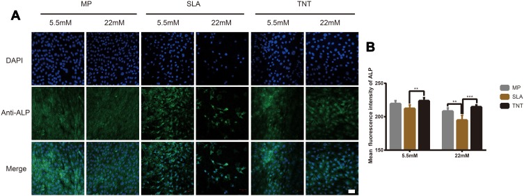
Figure 3.
High-glucose conditions inhibited ALP expression on different modified Ti surfaces. (A) The immunofluorescence staining of ALP observed by confocal scanning laser microscopy after osteogenic induction for 7 days. (B) Semi-quantitative analysis of the ALP immunofluorescence staining by Image J. High glucose levels inhibited ALP expression on different modified Ti surfaces, and TNT surface could alleviate the inhibition of ALP expression under high-glucose conditions. **p < 0.01, ***p < 0.001. Scale bar = 50 µm.

