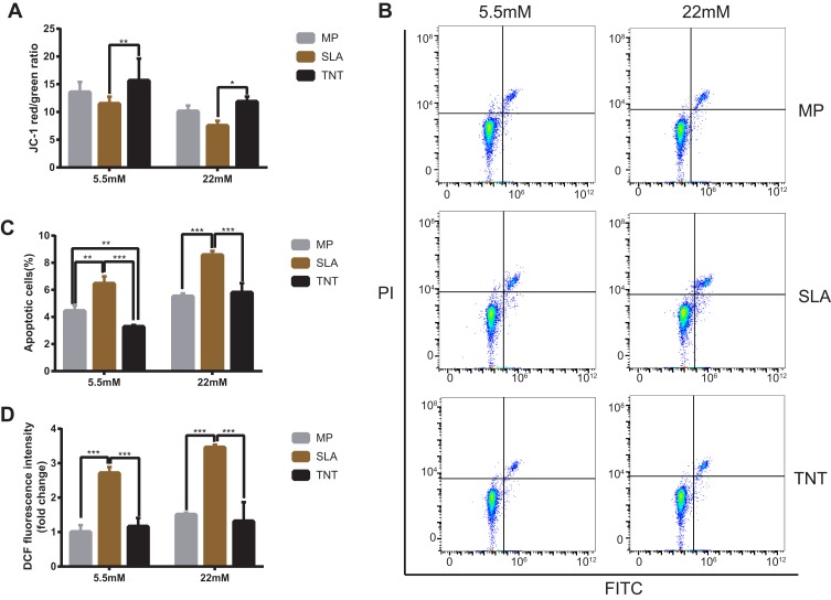
Figure 7.
High-glucose conditions decreased mitochondrial membrane potential, led to cell apoptosis, and induced excess intracellular ROS on different modified Ti surfaces. (A) The mitochondrial membrane potential determined by the red/green fluorescence intensity ratio in JC-1 staining. (B) Cell apoptosis detected with Annexin V–FITC/PI double-staining using flow cytometry. (C) Statistical analysis of the apoptosis rate on different modified Ti surfaces. Results showed that high-glucose conditions promoted cell apoptosis on different modified Ti surfaces. (D) Detection of intracellular ROS level on different modified Ti surfaces with the relative DCF fluorescence intensity as measured by flow cytometry. The average value of ROS in the MP-5.5-mM group is 1. Results showed that high-glucose conditions induced excess intracellular ROS level on different modified Ti surfaces, and the TNT surface could produce less oxidative stress than the SLA surface. *p < 0.05, **p < 0.01, ***p < 0.001. There was no significant difference between the two groups under normal or high-glucose conditions without *.

