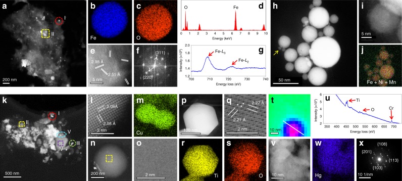
Fig. 2. Structural fingerprints of typical NPs extracted from human PE samples.
a HAADF-STEM image of the NPs extracted from the P4 sample. b–g Characterization of a spherical particle marked in the red circle in a (section I). b–c EDXS mapping, d EDXS spectrum, e atomic resolution HAADF-STEM image, f fast Fourier transform (FFT) image, and g EELS spectrum of the particle. h–j Characterization of the particles marked in the yellow square in a (section II). h HAADF-STEM image and j EDXS mapping of the particles in the section II in a. i, High-resolution HAADF-STEM image of a round particle marked by the yellow arrow in h. k, HAADF-STEM image of the particles extracted from the P27 sample. l Atomic resolution HAADF-STEM image and m EDXS mapping of particles marked in the red circle in k (section I). n HAADF-STEM image and o. High-resolution HAADF-STEM image of a spherical particle marked in the yellow square in k (section II). p HAADF-STEM image, q atomic resolution HAADF-STEM image, and r–s EDXS mapping of hexagonal particles marked in the green circle in k (section III). t EELS mapping of particles marked in the purple square in k (section IV) showing a core-shell structure of the particles. u EELS spectrum of the core part of the particle in t. v High-resolution HAADF-STEM image, w EDXS mapping, and x Selected area electron diffraction (SAED) pattern of the particles marked in cyan ellipse in k (section V). Source data for d, g, u are provided as a Source Data file.

