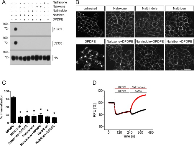
Figure 6.
Antagonist-selective inhibition of DPDPE-induced DOP receptor phosphorylation, internalization and G protein signaling. (A) Stably HA-hDOP receptor-expressing HEK293 cells were either not preincubated (−) or preincubated (+) with 5 µM naloxone, naltrindole, naltriben or naltrexone for 30 min at 37 °C, then stimulated with vehicle (water, -) or with 1 µM DPDPE (+) for 10 min at 37 °C. Cell lysates were then immunoblotted with antibodies to pT361 or pS363. Blots were stripped and reprobed with the anti-HA antibody. Blots are representative, n = 4. (B) Cells described in (A) were preincubated with anti-HA antibody and then treated with vehicle (DMSO), 5 µM naloxone, naltrindole or naltriben and with or without 1 µM DPDPE for 10 min at 37 °C. After fixation, cells were permeabilized, immunofluorescently stained and examined using confocal microscopy. Images are representative, n = 3. Scale bar, 20 µm. (C) Cells described in (A) were preincubated with anti-HA antibody and stimulated with vehicle (DMSO), 5 µM naloxone, naltrindole or naltriben and with or without 1 µM DPDPE for 10 min at 37 °C. Cells were then fixed and labeled with a peroxidase-conjugated secondary antibody. Receptor internalization was measured by ELISA and quantified as percentage of internalized receptors in agonist-treated cells. Data are means ± SEM from five independent experiments performed in quadruplicate. *p < 0.05 vs. DPDPE by one-way ANOVA with Bonferroni post-hoc test. (D) Reversal of DPDPE-induced hyperpolarization by naltrindole using a fluorescence-based membrane potential assay. After baseline recording for 60 sec, HA-hDOP receptor -expressing AtT-20 cells were exposed to 1 µM DPDPE and 240 sec later, 10 µM naltrindol was added, yielding a final molar DPDPE/antagonist ratio of 1:10. Shown are the normalized traces obtained from the average of four individual experiments performed in triplicates.

