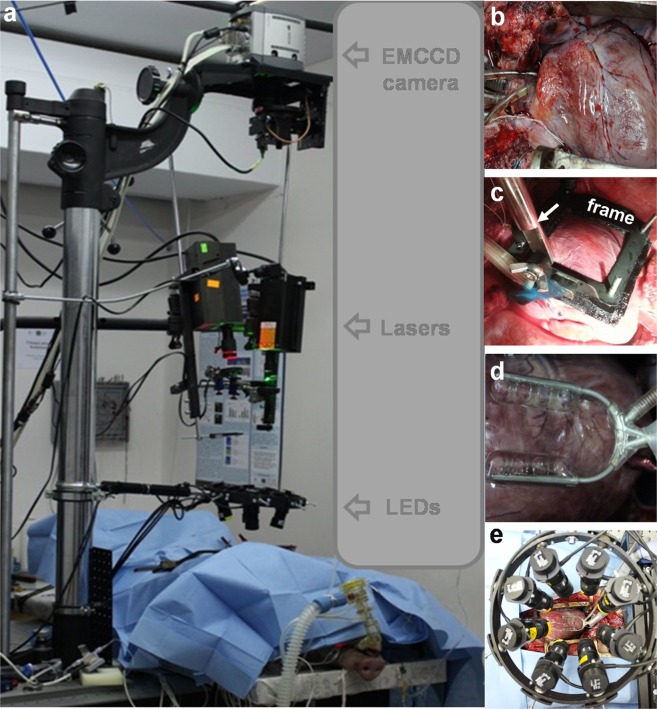
Figure 1.
Overall setup for optical mapping of the whole pig heart in situ. (a) Whole optical mapping setup adapted for registration of the cardiac electrical activity of the pig heart in situ during open-heart surgery. (b) Heart with inserted cannulas. (c) Hand-made frame for heart immobilization. Arrow indicates the bar. (d) Octopus tissue stabilizer used for heart immobilization. (e) Wheel with 8 positions for excitation LEDs (four 660 nm and four 780 nm).

