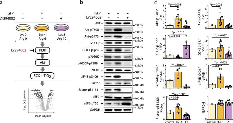
Fig. 1. Experimental setup to analyze PI3K/Akt signaling in contracting C2 myotubes.
a Contracting C2 myotubes, differentially labeled by stable isotopes using a triple stable isotope labeling with amino acids in cell culture (SILAC) approach and subjected to mild electrical pulse stimulation, were treated for 1 h with IGF-1 or LY294002 to stimulate or inhibit PI3K/Akt signaling as indicated. Cell lysates from triple SILAC experiments (n = 3 independent experiments) were individually subjected to SDS-PAGE and immunoblot analysis or mixed in equal amount for quantitative phosphoproteome analysis. Phosphopeptides were enriched using strong cation exchange chromatography and titanium dioxide chromatography (SCX-TiO2) and measured by LC-MS followed by computational data analysis. b Immunoblot analysis of PI3K/Akt/mTOR pathway activity using total and phospho-specific antibodies against established substrates of the canonical PI3K/Akt/mTOR pathway. c Quantification of immunoblot data from (b). Calculated signal intensities were normalized to the control and a two-tailed paired student’s t-test was performed. Error bars represent the SEM, n = 3-11 independent experiments.

