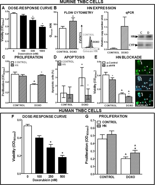
Figure 3.
Effect of HN on the chemosensitivity of TNBC cells. (A) Murine TNBC 4T1 cells were incubated in medium with FBS with liposomal Doxorubicin at different concentrations for 72 h (n = 6 replicates/condition). Viability was assessed by MTT assay. *p < 0.05 vs. control without Doxorubicin. ANOVA followed by Tukey’s test. (B) Mean fluorescence intensity (MFI) of HN in 4T1 cells as evaluated by flow cytometry (left panel) (n = 3 replicates/condition) and qPCR (right panel) following 24 h incubation with Doxorubicin (DOXO, 500 nM). Representative histogram and qPCR products are shown. *p < 0.05 Student’s t test. (C) 4T1 cells were incubated in medium with FBS with HN (10 μM) for 2 h before adding DOXO (500 nM) for further 72 h (n = 5 replicates/condition). Proliferation was assessed by BrdU incorporation (ELISA). ^p < 0.05 vs. respective control without DOXO; *p < 0.05 vs. respective control without HN. ANOVA followed by Tukey´s test. (D) 4T1 cells were incubated in medium with FBS with HN (10 μM) for 2 h before adding DOXO (500 nM) for further 24 h and apoptosis was evaluated by the TUNEL method. Bars indicate the percentage of apoptotic cells ± 95% confidence limits (CL) of the total number of cells counted in each specific condition (n ≥ 1000 cells/group). Confidence intervals for proportions were analyzed by the χ2 test: ^p < 0.05 vs. respective control without DOXO; *p < 0.05 vs. respective control without HN. χ2test. (E) 4T1 cells were transfected with p.Control or p.shHN and incubated with DOXO (500 nM) for 72 h (n = 6 replicates/condition). Viability was assessed by MTT assay. *p < 0.05 vs. p.Control, ^p < 0.05 vs. control without DOXO. ANOVA followed by Tukey´s test. Inset: Representative microphotograph showing positive cells for citrine (transfected cells, green, upper panel), nuclei stained with DAPI (middle panel) and their overlay (lower panel). Arrows indicate transfected cells. (F) MDA-MB 231 cells were incubated in medium with FBS containing liposomal Doxorubicin (DOXO) at different concentrations for 72 h (n = 6 replicates/condition). Viability was assessed by MTT assay. *p < 0.05 vs. control without DOXO. ANOVA followed by Tukey´s test. (G) MDA-MB 231 cells were incubated in medium with FBS and HN (10 μM) for 2 h before adding DOXO (250 nM) for further 72 h (n = 5 replicates/condition). Proliferation was assessed by BrdU incorporation (ELISA). ^p < 0.05 vs. respective control without DOXO, *p < 0.05 vs. respective control without HN. ANOVA followed by Tukey’s test.

