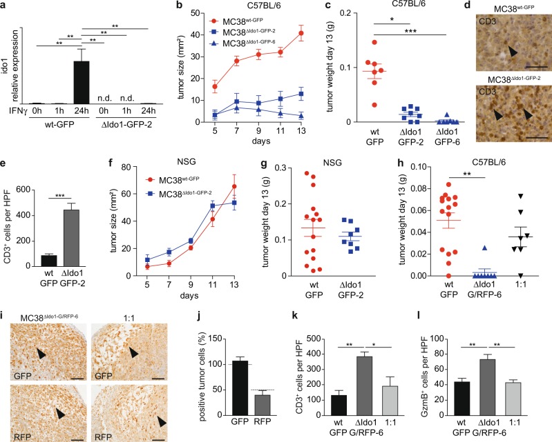
Fig. 4. Ablation of Ido1 in MC38 cells interferes with tumor formation in immunocompetent host mice.
a qPCR for Ido1 mRNA expression in MC38wt-GFP and MC38ΔIdo1-GFP-2 cells 0, 1, and 24 h after IFNγ stimulation (n = 3). b, c Tumor size (b) and final tumor weight (c) after subcutaneous injection of MC38wt-GFP, MC38ΔIdo1-GFP-2, and MC38ΔIdo1-GFP-6 cells into C57BL/6 hosts (MC38wt-GFP: 7 tumors, 7 host mice; MC38ΔIdo1-GFP-2: 7 tumors, 4 host mice; MC38ΔIdo1-GFP-6: 7 tumors, 7 host mice). d, e IHC staining (d) and quantification (e) of CD3+ infiltrating cells in tumors of MC38wt-GFP (7 tumors) and MC38ΔIdo1-GFP-2 (6 tumors) cells. f, g Tumor size (f) and final tumor weight (g) of MC38wt-GFP and MC38ΔIdo1-GFP-2 tumors in NSG hosts (MC38wt-GFP: 15 tumors, 15 host mice; MC38ΔIdo1-GFP-2: 8 tumors, 8 host mice). h Tumor weight of MC38wt-GFP (15 tumors, 15 host mice), MC38ΔIdo1-G/RFP-6 (8 tumors, 8 host mice), and 1 : 1 mixed tumors (7 tumors, 7 host mice) in C57BL/6 hosts. i, j IHC staining (i) and quantitation (j) of GFP+ and dsRed(RFP)+ tumor cells in mixed tumors (n = 3). Expected percentages of positive cells are indicated by dashed lines. Scale bars indicate 100 µm. k, l Quantification of CD3+ (k) and Granzyme B+ (l) immune cells in MC38GFP-wt (three tumors each), MC38ΔIdo1-G/RFP-6 (four tumors each), and mixed tumors (three tumors each). n.d.: not detectable. Bars represent mean ± SEM.

