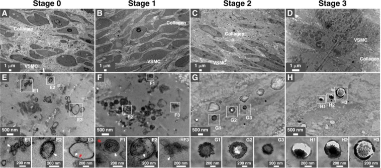
Figure 3.
Transmission electron microscopy observations of mineral particles in arteries of diabetic subjects. Artery tissues were prepared and observed under thin-section TEM as described in Methods. Low (A–D) and high (E–H) magnification images of tunica media are shown. Vesicles-like particles are denoted by arrows, with white arrows indicating single membranes and red arrows denoting double membranes. White rectangles indicate enlarged areas of the insets. VSMC, vascular smooth muscle cells.

