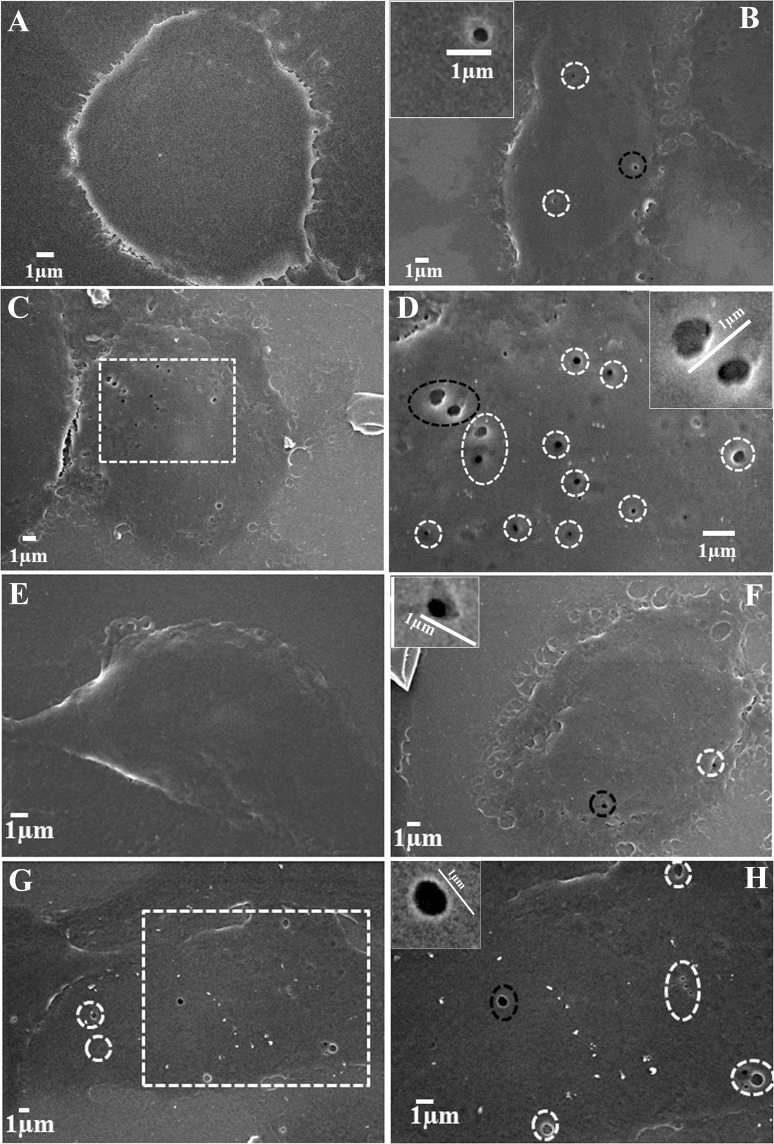
Figure 4.
Sonopores developed in the plasma membrane of cells after treatment with ultrasound in the presence and absence of MB (A) untreated MDA MB 231 cells (B) ultrasound treatment in absence of MB, dotted circles show pores. The black circled pore is shown in the inset (C) ultrasound treatment in the presence of MB, a magnified view of the dotted area is shown in the image (D). (D) Dotted circles surround pores and black circled pore is shown in the inset. B16F10 melanoma cells (E) untreated (F) treated with only ultrasound, (G) treated with ultrasound in presence of MB; the dotted area is magnified in image H, (H) dotted circles surround pores and black dotted circle pore is shown in the inset.

