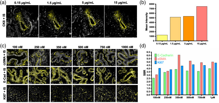
Fig. 1.
Ab-oligo conjugate and IS titration for optimal staining. (a) The CK8 Ab-oligo conjugate was titrated (0.15 to ) onto serial sections of normal breast FFPE tissue and equivalent concentrations of IS were applied to all tissue samples for visualization. Images are displayed with contrast and gain optimized for visualization of the positive CK8 staining pattern generated at each antibody conjugate concentration. (b) Image quantification showed the highest mean fluorescence intensity using antibody concentration for tissue staining. (c) IS was titrated (100 to 1000 nM) onto FFPE tissue with equivalent Ab-oligo conjugate concentrations present of -SMA (staining completed on normal breast tissue, top), E-Cad (staining completed on normal breast tissue, middle), and Ki-67 (staining completed on breast cancer tissue, bottom). (d) The SBR was calculated for each concentration tested for all biomarkers. The scale bars are displayed in all images.

