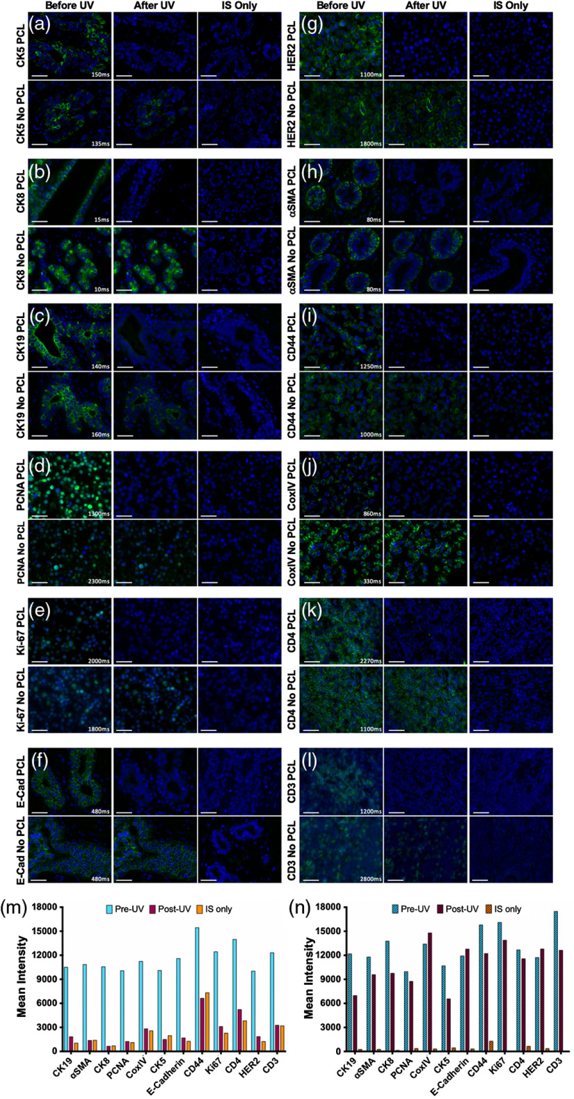
Fig. 4.
Ab-oligo signal removal validation using PCLs. Ab-oligo conjugate signal removal using PCLs was validated by staining serial sections using Ab-oligo conjugate () and IS without a PCL as well as Ab-oligo conjugate and IS with a PCL. Following image collection to demonstrate the Ab-oligo tissue staining pattern, each sample was treated for 15 min with UV light and images were again collected of each sample. Serial sections were stained with IS only and imaged as a negative control. The Ab-oligo staining pattern before and after UV treatment was verified for (a) CK5, (b) CK8, (c) CK19, (d) PCNA, (e) Ki67, (f) E-Cad, (g) HER2, (h) -SMA, (i) CD44, (j) CoxIV, (k) CD4, and (l) CD3. The mean fluorescence intensity for each image before and after UV light treatment was quantified for samples stained (m) with and (n) without a PCL. The scale bars are displayed in all images.

