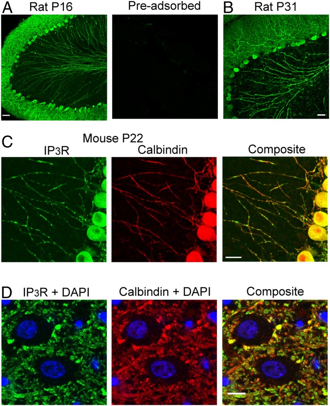
Fig. 1.
IP3R expression in PCs includes axons. (A and B) Low-magnification (20×) confocal immunofluorescence images of rat sagittal cerebellar slices stained with an antibody targeting type-I IP3Rs. Slices exposed to the same antibody preincubated with the immunogenic peptide were completely devoid of marker (A, Right). IP3R immunofluorescence, which is prominent in the somatodendritic compartment of PCs, clearly extends to the axons. Similar results were observed with young (A) and with adult rats (B). (Scale bar, 50 μm.) (C) The same expression pattern was present in mice; the assay included calbindin colabeling to highlight the colocalization of the two proteins along the axons. (Magnification, 63×.) (Scale bar, 20 μm.) (D) Zoom on a small field in the DCN highlights the presence of IP3R-I (Left) on synaptic-like boutons surrounding DCN somata, counterstained with DAPI. (D, Middle) PC axons and synaptic boutons labeled by calbindin. (D, Right) Composite for the three channels. (Scale bar, 5 μm.)

