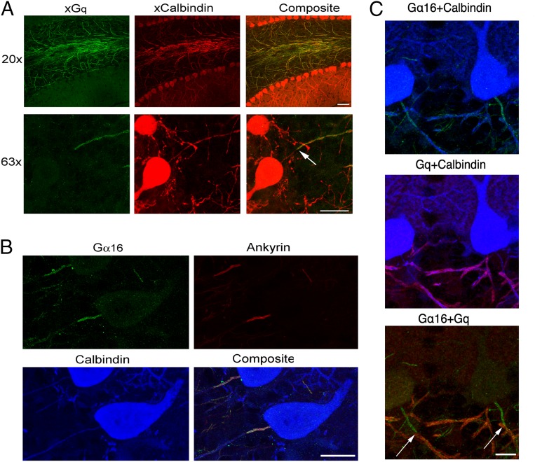
Fig. 4.
Expression of Gq class proteins in PC axons. (A, Top) Low-magnification immunofluorescence images using an antibody that targets Gq/α11 (green) show prominent Gq labeling along axons in the granule cell layer and diffuse labeling in the molecular layer; colocalization with calbindin (red) indicates that the axons belong to PCs. (A, Bottom) High-magnification images reveal that axonal Gq expression starts >20 μm away from the soma, leaving an initial stretch devoid of label (arrow). (B) Anti-Gα15/α16 antibody stains the proximal segment of PC axons. The spatial pattern of Gα15/α16 expression (green) coincides with that of ankyrin G (red; blue: calbindin counterstain marking PC outline). (C) Triple staining with calbindin (blue), Gα15/α16 (green), and Gq (red) showing complementary expression of the two proteins: Gα15/α16 at the initial segment, followed thereafter with no gap or overlap, by Gq expression in the distal portion (transitions at arrows). (Scale bars: A, Top: 50 μm; all others: 20 μm.)

