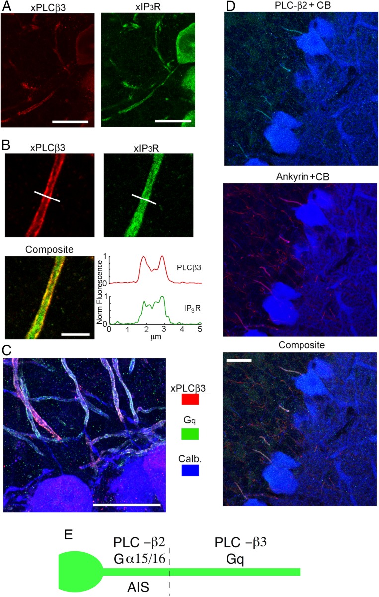
Fig. 5.
Complementary expression pattern of PLC-β2 and -β3 in PC axon. (A) Colabeling of PLC-β3 with IP3R. (A, Left) PLC-β3 is present in the soma and in the axon, but not in the initial segment. (A, Right) IP3R labeling is continuous. (Scale bar, 20 μm.) (B) PLC-β3 is membrane localized in the axon, whereas IP3Rs is distributed throughout the interior. Line profiles of the fluorescence intensity for the two markers (Bottom Right) highlight the different spatial patterns. (Scale bar, 5 μm.) (C) PLC-β3 and Gq exhibit a near-identical localization, both of them being excluded from the same stretch of proximal axon, in which calbindin labeling remains clearly visible. (Scale bar, 20 μm.) (D) Triple staining with antibodies against PLC-β2, calbindin, and ankyrin, showing that PLC-β2 is present exclusively in the initial segment where it costains with ankyrin. (Scale bar, 20 μm.) (E) Summary illustrating the differential localizations of Gα15/α16, Gq, PLC-β2, and PLC-β3.

