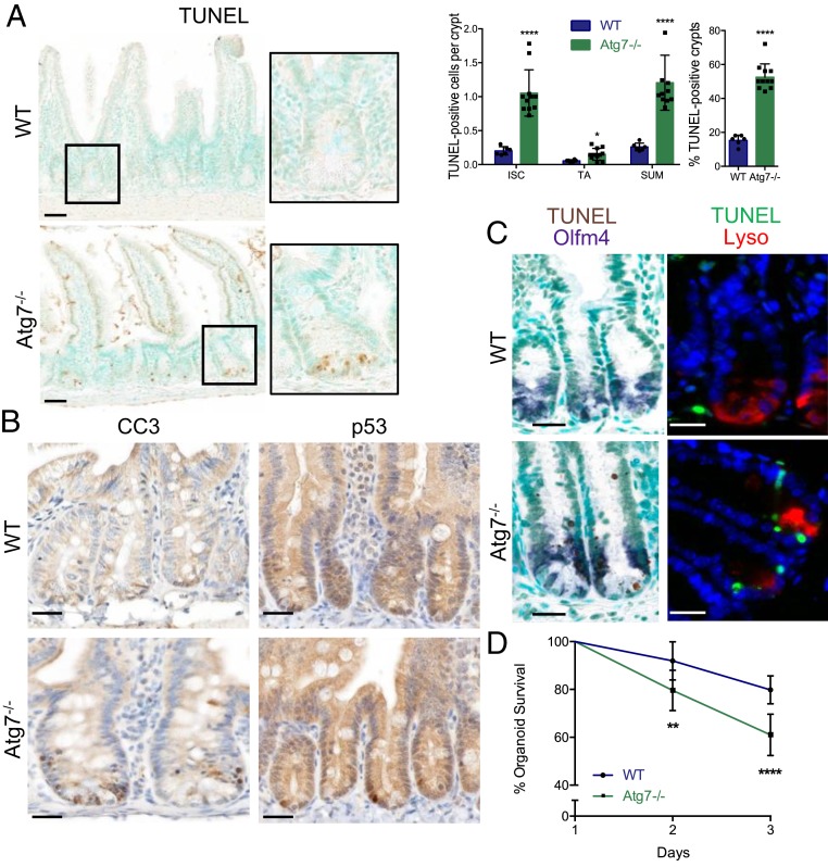
Fig. 1.
Atg7 deletion throughout the intestinal epithelium leads to Lgr5+ISC apoptosis. (A) Representative TUNEL staining on WT (wild type) and Atg7−/− tissue sections. Methyl green was used as a nuclear counterstain. Quantification of the percentage of TUNEL-positive crypts and the mean number of TUNEL-positive cells per crypt over 50 consecutive whole crypts in 6 WT and 11 Atg7−/− mice. The data shown are means ± SD. (Scale bars: 50 µm.) (B) Representative cleaved caspase-3 and p53 staining on tissue sections from WT and Atg7−/− intestines. (Scale bars: 25 µm.) (C) Representative TUNEL staining combined with in situ hybridization for Olfm4. Representative TUNEL staining combined with lysozyme staining. (Scale bars: 25 µm.) (D) Percentage survival from day 1 of organoids from the crypts of WT and Atg7−/− mice (n = 9 mice for WT and n = 7 mice for Atg7−/−). The data shown are means ± SD. Significant differences are shown with asterisks. *P < 0.05; **P < 0.01; ****P < 0.0001.

