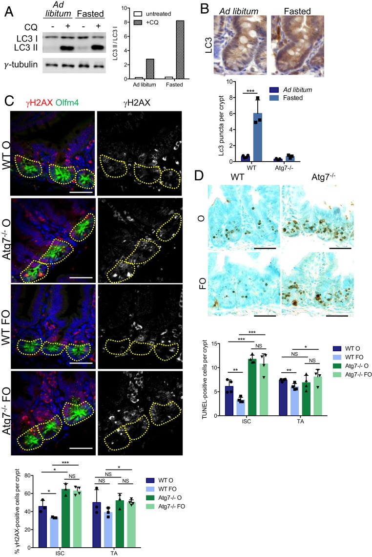
Fig. 4.
Fasting protects ISC from chemotherapy-induced DNA damage and apoptosis in an Atg7-dependent manner. (A) Western blotting for LC3 in organoids derived from WT mice that were either fed ad libitum or fasted for 24 h. Chloroquine (CQ) was used to block autophagic flux. γ-Tubulin was used as a loading control. (B) Representative LC3 staining in WT crypts from mice either fed ad libitum or fasted for 24 h. Quantification of LC3 puncta per crypt in WT and Atg7−/− mice either fed ad libitum or fasted for 24 h. (C) Representative z projection (from 20 stacks spanning 6 µm to include whole nuclei) of combined γH2AX and Olfm4 staining and γH2AX staining alone (Right) in the crypts of WT and Atg7−/− mice 6 h after oxaliplatin treatment alone (O) or preceded by a 24-h fast (FO). The percentage of γH2AX-positive cells was determined on at least 10 randomly selected whole crypts per mouse (n = 4 mice of each genotype). Cells with more than four γH2AX foci in their nuclei were considered γH2AX positive. Olfm4+ cells (circled area) were considered to be ISC, and the Olfm4− cells above them and below the crypt–villus junction were considered to be TA cells. The data shown are means ± SD. (Scale bars: 50 µm.) (D) Representative TUNEL staining of tissue sections from WT and Atg7−/− mice after 6 h of oxaliplatin treatment alone (O) or preceded by a 24-h fast (FO). Determination of the mean number of TUNEL-positive cells per crypt over 50 consecutive whole crypts in four mice per condition. The data shown are means ± SD. Significant differences are shown with asterisks. NS, not statistically significant. (Scale bars: 50 µm.) *P < 0.05; **P < 0.01; ***P < 0.005.

