Fig. 7.
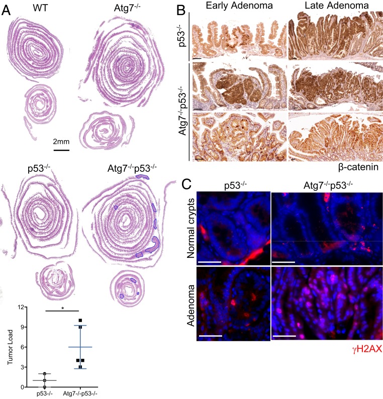
p53 blocks tumor initiation in the Atg7-deficient intestinal epithelium. (A) Representative hematoxylin and eosin staining of tissue sections of small intestinal and colonic “Swiss rolls” from WT, Atg7−/−, p53−/−, and Atg7−/−p53−/− mice killed 12 mo after tamoxifen treatment. Quantification of histologically assessed tumor load in p53−/− or Atg7−/−p53−/− mice 12 mo after tamoxifen treatment (n = 3 p53−/− and n = 5 Atg7−/−p53−/− mice). Tumors are circled in blue. (Scale bar: 2 mm. Significant differences are shown with asterisks. *P < 0.05.) (B) Representative β-catenin staining on small polyps (Left) and adenomas (Right) from p53−/− and Atg7−/−p53−/− mice killed 12 mo after tamoxifen treatment. (Scale bars: 50 µm.) (C) Representative γH2AX staining on healthy crypts and adenomas from p53−/− and Atg7−/−p53−/− mice killed 12 mo after tamoxifen treatment. (Scale bars: 50 µm.)

