Fig. 5.
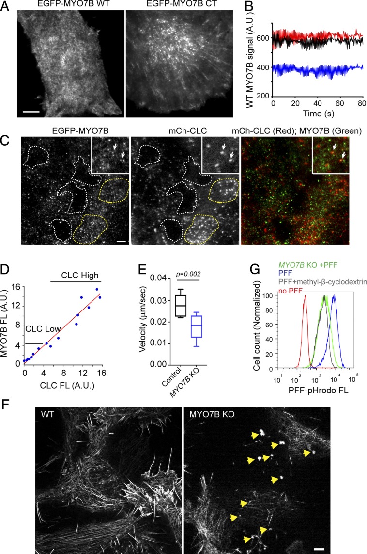
MYO7B maintains plasma membrane dynamics at domains enriched in clathrin. (A) TIRF microscopy analysis of plasma membrane binding by EGFP-WT MYO7B and EGFP-CT MYO7B in cells stably expressing low levels of these proteins (Scale bar: 5 µm.) (B) Quantification of representative EGFP-WT MYO7B signal on the plasma membrane in a time-lapse video. Shown are fluorescence intensities of three randomly chosen spots measured by Fiji. A.U., arbitrary unit. (C and D) MYO7B binds to the plasma membrane in clathrin-enriched domains. (C) TIRF microscopy analysis of cells stably expressing EGFP-WT MYO7B and mCh-CLC. (Insets) Close-up views of cells revealing limited colocalization between MYO7B and CLC. The dashed lines indicate surface regions either lacking clathrin (white) or enriched in clathrin (yellow). (D) Correlation between CLC and MYO7B fluorescence (FL) intensity in cell surface domains as outlined in Fig. 5C (R2 = 0.956 by linear regression). (E) MYO7B inactivation affects plasma membrane dynamics. Control and MYO7B-KO cells were stained with GFP+ and imaged by time-lapse confocal microscopy (SI Appendix, Fig. S5C and Movies S3 and S4). The whisker graph shows the plasma membrane ruffling velocity determined by the Nikon Element analysis of videos taken from three independent experiments. Control, n = 6 cells; MYO7B-KO, n = 12 cells. P values by two-tailed unpaired Student’s t test. (F) MYO7B-KO cells have reduced actin fibers under the plasma membrane. TIRF-SIM analysis of WT or MYO7B-KO cells transfected with EGFP-tractin. Note the presence of actin-containing aggregates in MYO7B-KO cells (indicated by arrows) (Scale bar: 10 µm.) (G) Depletion of cholesterol inhibits α-Syn PFF endocytosis. Cells were treated with methyl-β-cyclodextrin (5 mM) or DMSO for 30 min before incubation with α-Syn PFF pHrodo and FACS analysis.

