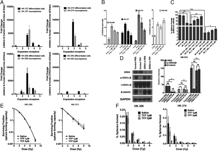
Fig. 2.
TFP prevents radiation-induced phenotype conversion in GBM. (A) Four primary patient-derived GBM lines (HK-157, HK-374, HK-308, and HK-345) were grown as differentiated cells in the presence of 10% serum and as gliomaspheres in serum-free medium. RNA from both cultures was isolated, and qPCR was performed to analyze for the difference in dopamine receptor (DRD1, DRD2, DRD3, DRD4, and DRD5) expression levels. GAPDH was used as the internal control to obtain the dCt (delta Ct) values. The dCt values were normalized to DRD5 across all lines to obtain the ddCt values. The fold change in expression levels of DRDs was calculated by 2^-ddCt method. (B) Sorted ZsGreen-cODC−negative cells from HK-308, HK-157, HK-374, and HK-345 ZsGreen-cODC vector-expressing cells were plated at a density of 50,000 cells per well in a six-well plate and, the following day, pretreated either with TFP (1 and 5 μM) or vehicle (saline) 1 h before irradiation at a single dose of 0 or 8 Gy. Five days later, the cells were detached and analyzed for ZsGreen-cODC−positive cells by flow cytometry, using their noninfected parent lines as controls, and represented as percentage iGICs. (C) Sphere formation assay was performed using the HK-308 parent gliomaspheres plated in a 96-well plate and treating them with different concentrations of TFP (1 and 5 μM) in combination with or without bromocriptine (0.1, 1, and 5 μM). The spheres were fed with growth factors (10×) medium regularly. The number of spheres formed in each condition was counted and normalized against the vehicle control. The resulting data were presented as percentage spheres formed. (D) HK-374 cells were treated with either control siRNA or DRD2-specific siRNA for 72 h, serum-starved for 4 h, and then treated with or without TFP (10 μM). Proteins were extracted and subjected to Western blotting. The membranes were blotted for p-GSK3a/b, t-GSK3a/b, β-catenin, and GAPDH. The intensity of each band was calibrated using ImageJ and presented as density ratio of gene over GAPDH in HK-374 cells. (E and F) Parent HK-308 and HK-374 gliomaspheres seeded in a 96-well plate were pretreated either with TFP (1 or 5 μM) or vehicle (saline) 1 h before irradiation at a single dose of 0, 2, 4, 6, or 8 Gy. This setup was maintained with regular feeding of the spheres with 10× growth factors. The number of spheres formed in each condition was counted and normalized against the respective nonirradiated control. The final curve was generated using a linear quadratic model. All experiments in this figure have been performed with at least three biological independent repeats. P values were calculated using unpaired t test. *P value < 0.05, **P value < 0.01, and ***P value < 0.001.

