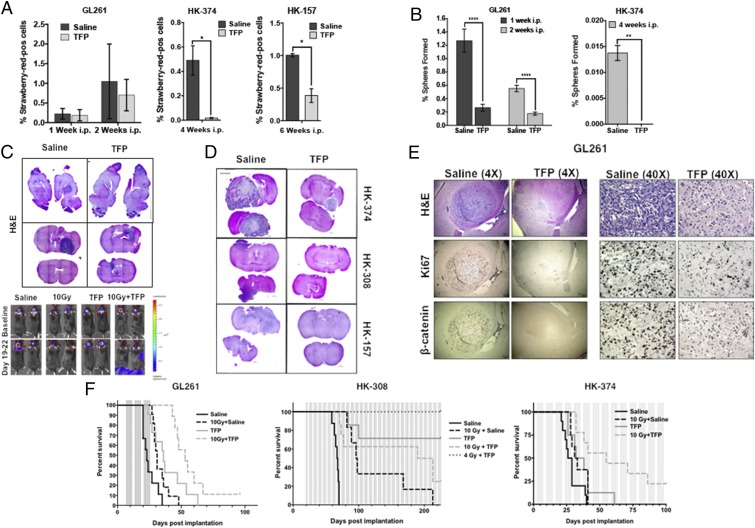
Fig. 6.
TFP in combination with radiation prolongs survival in mouse models of GBM. (A) The 2 × 10⁵ GL261-StrawRed cells were implanted into the brains of the C57BL/6 mice, and 3 × 10⁵ HK-374 and HK-157-StrawRed cells were implanted into the brains of NSG mice. Tumors were grown for 3 d (HK-374) and 7 d (HK-157 and GL261) for successful grafting. Mice bearing tumors were injected i.p. on a 5-d on/2-d off schedule for 1 or 2 wk (GL261), 4 wk (HK-374), and 6 wk (HK-157) either with TFP (20 mg/kg) or saline. At the end of the treatment, brains of these mice were extracted, and tumors were digested and sorted for StrawRed cells. The data from the sort were plotted and presented as percentage of StrawRed-positive cells of the total number of cells in each GBM line. (B) The sorted StrawRed cells from A were plated on 96-well plates in serum-free medium and fed with fresh growth factors every 2 d to 3 d. The number of spheres formed in each condition was counted and normalized against the respective control. (C) Hematoxylin/eosin (H&E) stained sagittal and coronal sections of the brains of C57BL/6 mice intracranially implanted with 2 × 10⁵ GL261 cells. Tumors were grown for 7 d for successful grafting. Mice were injected with TFP or saline i.p. on a 5-d on/2-d off schedule for 3 wk. TFP treatment led to a marked reduction in tumor size. (D) H&E stained coronal sections of the NSG mice brains implanted with HK-374, HK-308, and HK-157-GFP-Luc cells which were treated continuously with either TFP (s.c., 20 mg/kg) or saline until they met the criteria for study endpoint. (E) The 4× and 40× images of coronal sections of brains from C57BL/6 mice implanted with GL261-GFP-Luc cells intracranially and treated with either TFP (20 mg/kg) or saline i.p. for 2 wk and stained for H&E, Ki67, and β-catenin. (F) Survival curves for C57BL/6 mice (Left) implanted intracranially with 2 × 10⁵ GL261-GFP-Luc cells and NSG mice injected intracranially with 3 × 10⁵ HK-308-Luc (Middle) or HK-374-GFP-Luc (Right) cells. Tumors were grown for 3 d (HK-374), 7 d (GL261), and 21 d (HK-308) for successful grafting. Mice were injected with either TFP (20 mg/kg) or saline i.p. for GL261 for 3 wk, and s.c. for HK-308 and HK-374 lines, continuously until they reached the study endpoint. Log-rank (Mantel−Cox) test for comparison of Kaplan−Meier survival curves indicated significant differences in the TFP-treated mice compared to their respective controls. P values in GL261 study: saline vs. TFP (***P value = 0.00014), saline vs. 10 Gy + saline (*P value = 0.0126), saline vs. 10 Gy + TFP (***P value <0.0002), TFP vs. 10 Gy (P value = 0.5320), TFP vs. 10 Gy + TFP (*P value = 0.0324). P values in HK-308 study: saline vs. TFP (***P value = 0.00014), saline vs. 10 Gy + saline (***P value = 0.0004), saline vs. 10 Gy + TFP (****P value 0.0001), saline vs. 4 Gy + TFP (**P value = 0.0011), TFP vs. 10 Gy (*P value = 0.0141), TFP vs. 10 Gy + TFP (P value = 0.1052), TFP vs. 4 Gy + TFP (P value = 0.2137). P values in HK-374 study: saline vs. TFP (*P value = 0.0361), saline vs. 10 Gy + saline (*P value = 0.0413), saline vs. 10 Gy + TFP (***P value = 0.00025), TFP vs. 10 Gy (P value = 0.7217), TFP vs. 10 Gy + TFP (*P value = 0.0273). GBM-StrawRed experiments in this figure have been performed with at least two biological repeats. P values were calculated using unpaired t test. *P value < 0.05, **P value < 0.01, and ****P value < 0.0001.

