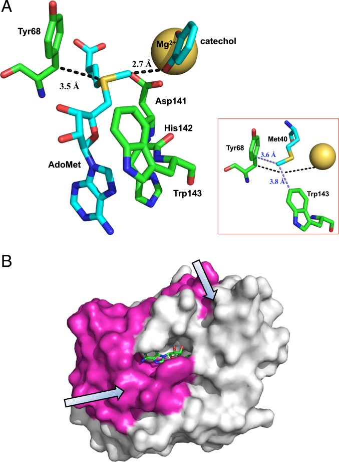
Fig. 7.
A model for the COMT reaction integrating the HDX and kinetic findings. (A) The active site of COMT, illustrating the proposed duality of roles for Tyr68 in controlling the methyl donor–acceptor distance along the reaction coordinate (dashed black line) and the sampling of multiple conformational substates involving side chains orthogonal to the reaction coordinate (dashed light blue line, Inset). Structure is based on PDB ID code 3BWM (36), not showing dinitro groups on the catechol. (B) Space filling model of COMT showing the dynamical regions of the protein that extend in orthogonal directions from the protein solvent interface toward the reacting atoms in the active site.

