Fig. 6.
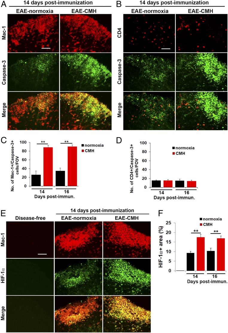
CMH enhances apoptosis and HIF-1α expression in monocytes. (A, B, and E). Frozen sections of lumbar spinal cord taken from disease-free, EAE–normoxia or EAE–CMH mice at the peak phase of disease (14 d postimmunization) were stained for Mac-1 (Cy-3) and cleaved caspase-3 (AlexaFluor-488) in A, CD4 (Cy-3) and cleaved caspase-3 (AlexaFluor-488) in B, and Mac-1 (Cy-3) and HIF-1α (AlexaFluor-488) in E. (Scale bar, 25 μm.) (C, D and F). Quantification of the number of Mac-1/cleaved caspase-3 dual-positive (C) or CD4/cleaved caspase-3 dual-positive (D) cells/FOV or HIF-1α+ area (F). Results are expressed as the mean ± SEM (n = 6 mice per group). Note that during the peak phase of disease (days 14–16), CMH enhanced the death of infiltrated monocytes and increased HIF-1α expression in these cells. **P < 0.01.

